Health & Living
Muscular tissue has one major purpose: to convert ATP into motion. All three types of muscular tissue — skeletal, cardiac, and smooth — are specialized for this sole purpose. The muscular system is an organ system that utilizes all types of muscular tissue, and concentrates on maintaining postural support and producing movements of the bones.
Muscles have five main functions:
-
Movement: muscles not only move our body from place to place, but also internal body contents, aiding in blood circulation, breathing, and digestion.
-
Stability: muscles stabilize our body by preventing any unwanted movements, thus maintaining posture.
-
Control of body openings and passages: Muscles often encircle various parts of the body to regulate and maintain homeostasis. Sphincters, for example, are heavily involved in elimination of waste.
-
Heat production: Skeletal muscles produce the majority of body heat, which is important in metabolism.
-
Glycemic control: Skeletal muscles play a large role in the regulation of blood glucose concentration, as they absorb, store, and use the body’s glucose.

Muscle Anatomy
Skeletal muscle is not only composed of muscular tissue, but also connective tissues, nerves, and blood vessels. There are four connective tissues of skeletal muscle:
-
Endomysium is the smallest and deepest. It is comprised of a thin sleeve of loose connective tissue that surrounds each muscle fiber. By creating spaces for blood capillaries and nerve fibers to encompass the fibers, all fibers are stimulated and nourished.
-
Perimysium wraps muscles fibers together to form a fascicle using a thicker connective tissue. It carries larger vessels and nerves, as well as muscle spindles.
-
Epimysium is a fibrous sheath surrounding the entire muscle.
-
Fascia is the largest and most superficial. It is a sheet of connective tissue that separates muscles from each other and from the subcutaneous tissue.

Muscle shape and compartmentalization vary greatly depending on its function. Depending on the muscle’s strength and direction of pull, the fascicular orientation can vary greatly. There are five types of muscles classified by fascicle orientation: fusiform, parallel, triangular, pennate, and circular.

Muscle compartmentalization is when similar-functioning muscles are enclosed in close proximity. Each compartment also contains the nerves and blood vessels supplying the muscle group.
There are two types of muscle attachments: direct and indirect. In direct attachments, the muscle is so close to the bone that the tissue seems to emerge straight from the bone. In indirect attachments, there are gaps between the muscle and bone attachment, which are filled with tendon, a fibrous band or sheet.
You can tell what a muscle does by its origins and insertions. An origin is where the muscle attaches and is stationary. An insertion is where the muscle attaches and is mobile, producing movement. The biceps brachii, for example, originates at the scapula and inserts onto the radius. It thus produces elbow flexion.

Based on a muscles ability to produce or prevent movement, it can be placed into a certain functional group:
-
The agonist muscle produces the majority of the force during a joint action. The brachialis, for example, is the prime mover in elbow flexion.
-
The synergist muscle aids the prime mover. The biceps brachii works with the brachialis to flex the elbow.
-
The antagonist muscle works against the agonist muscle, oftentimes providing tension so as to prevent oneself from excessive movement. The triceps brachii extends the elbow, the opposite movement of elbow flexion.
-
The fixator muscle stops a bone from moving, holding it steady for another muscle to pull. The rhomboid muscles are, like the biceps brachii, attached at the scapula. When contracted the rhomboid holds the scapula in place so the force is exerted on the radius instead.

Muscles of the Head and Neck
Muscles of the Head and Neck will be divided into three categories:
-
Muscles of Facial Expression
-
Muscles of Chewing and Swallowing
-
Muscles that Move the Head
Muscles of Facial Expression
Humans have more expressive faces than other mammals or animals due to our multitude of facial muscles. These muscles can tense or relax the skin to produce smiles or frowns, as well as contributing to speech or mastication. Important muscles include the frontalis, occipitalis, orbicularis oculi, orbicularis oris, mentalis, buccinator, and platysma.

Muscles of Chewing and Swallowing
Many muscles aid in food manipulation with tongue movements, chewing, and swallowing. These muscles include the temporalis, masseter, pterygoids, hyoid muscles, and pharyngeal constrictors.

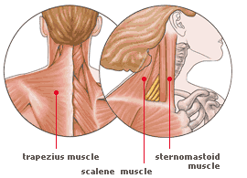
Muscles that Move the Head
Muscles involved in head movement do so by flexion, extension, hyperextension, and rotation. Many head actions actually involve a combination of these movements, such as looking over your shoulder. Important muscles include the sternocleidomastoid, scalenes, and trapezius.
Muscles of the Trunk
Muscles of the trunk will be divided into three function groups:
-
Respiration
-
Abdominal wall
-
Vertebral column movement
Respiration
The diaphragm and intercostal muscles are the primary reason why we are able to breathe. The contraction of the diaphragm enlarges the thoracic cavity, causing inspiration, while its relaxation causes expiration as the diaphragm shrinks the thoracic cavity. The intercostals stiffen the thoracic cage, allowing rigidity and support so as to prevent the cage from collapsing inward.

Vertebral Column Movement
Muscles of the back aid primarily in extension, rotation, and flexion. Important muscles include the latissimus dorsi, trapezius, iliocostalis, spinalis, and longissimus.
Muscles Acting on the Upper Limb
Muscles acting on the shoulder
The scapula is capable of many movements, such as rotation, elevation, depression, protraction, and retraction. Muscles include the pectoralis minor, serrates anterior, trapezius, levator scapulae, rhomboid minor, and rhomboid major.

Abdominal wall
Although the abdominal cavity has little skeletal support, it is enclosed with various muscles to strengthen the abdominal wall. These muscles include the external abdominal obliques, internal abdominal obliques, transverse abdominals, and rectus abdominus.


Muscles acting on the arm
Many muscles are involved in arm movement, including the pectoralis major, latissimus dorsi, deltoid, teres major, and rotator cuff muscles.

Muscles acting on the forearm
The forearm and elbow performs flexion, extension, pronation and supination. Muscles that perform these movements include the brachialis, biceps brachii, triceps brachii, brachioradialis, pronator teres, and supinator.

Muscles acting on the wrist and hand
These muscles mainly produce wrist and finger flexing and extending, as well as radial and ulnar flexion, and finger abduction and adduction. These muscles include the flexor carpi radialis, flexor carpi ulnaris, flexor digitorum superficialis, palmaris longus, flexor pollicis longus, extensor carpi radialis longus, extensor carpi radialis brevis, extensor digitorum, extensor carpi ulnaris, abductor pollicis longus, extensor pollicis brevis, and extensor pollicis longus.

Muscles Acting on the Hip and Lower Limb
Muscles acting on the Hip and Femur
Muscles that aid in hip and femur movement include the iliacus, psoas major, tensor fasciae latae, gluteus maximus, gluteus medius, gluteus minimus, quadratus femoris, adductor brevis, adductor longus, gracilis, and pectineus.

Muscles acting on the knee and leg
These muscles form the thigh and thus perform actions on the knee joint. This includes the quadriceps femoris, rectus femoris, vastus lateralis, vastus medialis, vastus intermedius, sartorius, biceps femoris, semitendinosus, semimembranosus, and popliteus.

Muscles acting on the foot
The muscles that provide foot movement are similar to those in the hands. They include the extensor digitorum longus, extensor hallucis longus, tibialis anterior, gastrocnemius, soleus, flexor digitorium longus, flexor hallucis longus, fibularis brevis, and fibularis longus.

Using Anatomy to Build Muscle
The quadriceps are composed of the vastus lateralis, vastus medialis, vastus intermedius, and rectus femoris. In conjunction, these four muscles work together to extend the knee. In order to enhance the shape of your quads, do moves such as squats and leg extensions! These exercises work on knee extension, and will thus produce larger, stronger quadricep muscles, thus creating the coveted outer-thigh sweep of the vastus lateralis and teardrop shape of the inner thigh, which comes from the vastus medialis.
